
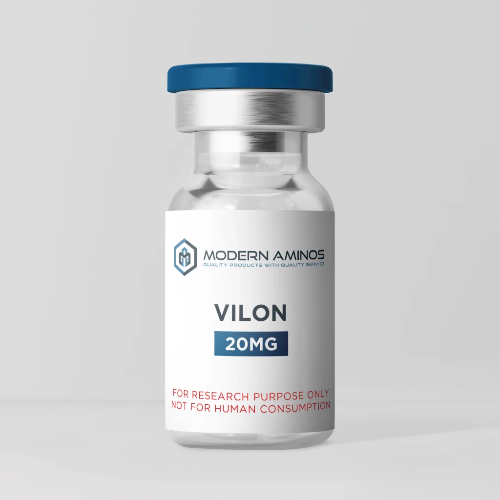
What Is Vilon?
Vilon is the dipeptide Lys-Glu, written KE in one-letter code: one L-Lysine joined to one L-Glutamic acid. PubChem lists it as CID 7010502, with the formula C11H21N3O5 and a molecular weight of 275.30 g/mol. Unlike most short bioregulators, it has measured data at PEPT1, the transporter built for di- and tripeptides.
Chemical Identity of Vilon
Picture two beads on a string. The first bead, L-Lysine, has a long, flexible four-carbon arm that ends in an amine. The second bead, L-Glutamic acid, has a shorter two-carbon arm that ends in a carboxylic acid. Add the amine at the very front of the chain and the acid at the very end, and you have two amines and two acids.
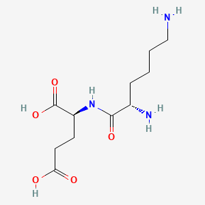
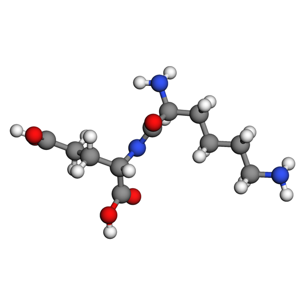
That balance matters. Near neutral pH, amines tend to pick up a proton and acids tend to give one up, so Lys-Glu carries plus and minus charges in roughly equal measure. Chemists call that a zwitterion. Reasonable inference: with only one positively charged side chain, Lys-Glu does not fit the doubly positive pattern that PEPT1 refused to carry in the 2006 study, which is consistent with its measured transport. Both stereocenters sit in the L, or S, configuration, and the InChIKey UGTZHPSKYRIGRJ-YUMQZZPRSA-N encodes that handedness.
Molecular Data
| Property | Value |
|---|---|
| CAS# | 45234-02-4 |
| PubChem ID | CID 7010502 |
| Molecular Formula | C11H21N3O5 |
| Molecular Weight | 275.30 g/mol |
| Exact Mass | 275.1481 Da |
| IUPAC Name | (2S)-2-[[(2S)-2,6-diaminohexanoyl]amino]pentanedioic acid |
| InChIKey | UGTZHPSKYRIGRJ-YUMQZZPRSA-N |
| SMILES | C(CCN)C[C@@H](C(=O)N[C@@H](CCC(=O)O)C(=O)O)N |
| Molecular Class | Dipeptide (Lys-Glu); short bioregulator peptide |
| Stereocenters | 2 defined, 0 undefined |
| Hydrogen bond donors / acceptors | 5 / 7 |
| XLogP3 (computed) | -6.3 |
Values from PubChem CID 7010502 (computed by PubChem).
Hello, neighbor. Today’s molecule is about as small as a peptide can be. Vilon is just two amino acids holding hands. But small things can teach big lessons, and this little pair has a story that most of its larger cousins do not: someone actually measured how it behaves at a real transporter. So let’s walk through that story one step at a time.
Peptide Transporters: The Research Picture
First, a little background. PEPT1, also called SLC15A1, is a membrane protein whose job is to move di- and tripeptides across a membrane, using the energy of protons moving along with them. Chemists who study it ask two separate questions. Does a molecule stick to the transporter? And does the transporter actually carry it across? Those sound like the same question, but they are not, and Vilon helps show why.
Step 1: Recognition at PEPT1. To be carried, a molecule first has to be recognized. In a 2005 modeling study of PEPT1 substrates, Lys-Glu is recorded with an affinity constant (Ki) of 1.3 mM. A year later, a pharmacophore study measured how well it blocks the uptake of Gly-Sar, the standard PEPT1 reference dipeptide, and found an IC50 of 0.82 mM. Two labs, two methods, and both land in the low-millimolar range.
Step 2: From binding to transport. The same 2006 study then asked whether PEPT1 actually moves each peptide, using a membrane depolarization assay. Lys-Glu produced a transport signal with an EC50 of 0.53 mM and reached 121% of the Gly-Sar maximum. That makes it a true substrate, not just a binder. The authors also found that dipeptides carrying two positive charges were not carried at all.
Step 3: The docking view. In 2023, a team led by Khavinson and Petukhov docked 26 short peptides into computer models of four transporters. Lys-Glu scored −19.02 at LAT1, −23.94 at LAT2 and −17.02 at PEPT1. The ICM-Score is a partial analogue of binding free energy, so more negative numbers mean a stronger predicted fit. At LAT2, Lys-Glu ranked 10th of 26 and beat the reference inhibitor BCH, which scored −15.15.
Step 4: A predicted DNA address. A 2016 docking study built models of DNA-peptide complexes for 19 short peptides. It placed Lys-Glu, along with Glu-Asp-Pro, at the four-base sequence agat. A separate 2020 survey counted the KE motif across 20,417 UniProt protein sequences and found it most often in cytoplasmic and nuclear proteins.
Step 5: What the numbers imply for assay design. Reasonable inference: because the measured PEPT1 values sit near 1 mM, Lys-Glu is best compared with Gly-Sar at matched millimolar concentrations, while the docking scores are useful for ranking it against relatives such as Lys-Glu-Asp. A measured EC50 and a docking score answer different questions, and they should be reported side by side, not swapped for each other.
Assay and Identity Data
| Item | Detail | Source |
|---|---|---|
| Sequence | H-Lys-Glu-OH (KE) | PubChem, ChEBI |
| CAS | 45234-02-4 | PubChem (synonym) |
| PubChem CID / ChEBI | 7010502 / CHEBI:74557 | PubChem, ChEBI |
| Formula | C11H21N3O5 | PubChem |
| Molecular weight | 275.30 g/mol (exact mass 275.1481 Da) | PubChem |
| XLogP / TPSA | −6.3 / 156 Ų | PubChem |
| PEPT1 Ki | 1.3 mM | Biegel et al., 2005 |
| PEPT1 Gly-Sar uptake IC50 | 0.82 mM | Vig et al., 2006 |
| PEPT1 transport EC50 | 0.53 mM (121% of Gly-Sar maximum) | Vig et al., 2006 |
| ICM-Score, LAT1 / LAT2 / PEPT1 | −19.02 / −23.94 / −17.02 | Khavinson et al., 2023 |
| Predicted DNA docking site | agat | Khavinson et al., 2016 |
The measured rows and the modeled rows agree on the big picture. Lys-Glu is recognized by PEPT1 and carried by it, at low-millimolar values. The docking rows add a ranking, and they place Lys-Glu nearer the middle of the pack at LAT2 and lower at LAT1 and PEPT1. An XLogP of −6.3 describes a molecule that strongly prefers water to oil, which is just what you would expect from two amines and two acids.
Why Not Livagen for the Same Question?
Here is a fair question. Livagen is Lys-Glu-Asp-Ala, the same KE start with two more residues. Why not use it? For PEPT1 work, length is the answer. The 2006 study found that tetrapeptides could neither bind to nor be carried by PEPT1, so a dipeptide like Vilon is the right size for measured PEPT1 questions. Livagen’s PEPT1 value, −16.70, comes only from docking. For questions about how added residues change predicted transporter binding, though, comparing Vilon with Livagen or with Prostamax (Lys-Glu-Asp-Pro, −33.42 at PEPT1) is exactly the right move.
Research Supply and Documentation
All applicable tests are performed on our products. Modern Aminos supplies Vilon 20mg as a white lyophilized powder in a single vial format. The current Freedom Diagnostics Testing certificate lists mass identification as Vilon and reports purity by HPLC with UV detection coupled with mass spectrometry, along with endotoxin results. Certificates can be looked up on the COA Search page, and our explainer on why HPLC testing matters walks through the purity method one step at a time.
Related Research Tools
References
- Biegel A, Gebauer S, Hartrodt B, Brandsch M, Neubert K, Thondorf I. Three-dimensional quantitative structure-activity relationship analyses of beta-lactam antibiotics and tripeptides as substrates of the mammalian H+/peptide cotransporter PEPT1. J Med Chem. 2005;48(13):4410-4419. PubMed 15974593
- Vig BS, Stouch TR, Timoszyk JK, et al. Human PEPT1 pharmacophore distinguishes between dipeptide transport and binding. J Med Chem. 2006;49(12):3636-3644. PubMed 16759105
- Khavinson VK, Lin’kova NS, Tarnovskaya SI. Short Peptides Regulate Gene Expression. Bull Exp Biol Med. 2016;162(2):288-292. PubMed 27909961
- Terekhov AY, Kormilets DY, Linkova NS, Kuznik BI, Mar’yanovich AT, Khavinson VK. Peptide KE in Human Proteome. Bull Exp Biol Med. 2020;168(5):631-633. PubMed 32246368
- Khavinson VK, Linkova NS, Rudskoy AI, Petukhov MG. Feasibility of Transport of 26 Biologically Active Ultrashort Peptides via LAT and PEPT Family Transporters. Biomolecules. 2023;13(3):552. PMC10046148
- National Center for Biotechnology Information. PubChem Compound Summary for CID 7010502, Lysylglutamic acid (Vilon). pubchem.ncbi.nlm.nih.gov/compound/7010502
- European Bioinformatics Institute. ChEBI entry CHEBI:74557, Lys-Glu. ebi.ac.uk/chebi/CHEBI:74557
Frequently Asked Questions
What Is Vilon?
Vilon is a synthetic dipeptide made of two amino acids in the order Lys-Glu, written KE in one-letter code. It belongs to the Khavinson family of short peptide bioregulators, and PubChem lists it as CID 7010502 (CAS 45234-02-4), C11H21N3O5, 275.30 g/mol.
Is Vilon a Peptide?
Yes. Vilon is two L-amino acids, L-Lysine and L-Glutamic acid, joined by one peptide bond. Because it has two residues, chemists call it a dipeptide, the shortest kind of peptide.
How Does Vilon Work?
Its best-documented target is the PEPT1 peptide transporter. Lys-Glu binds PEPT1 with a Ki of 1.3 mM and is transported with an EC50 of 0.53 mM. Docking models add predicted binding at the LAT1 and LAT2 transporters and at the DNA sequence agat.
Vilon vs. Livagen: What Is the Difference?
Two residues. Livagen is Lys-Glu-Asp-Ala, so it is Vilon plus Aspartic acid and Alanine. That length matters for PEPT1, which handles di- and tripeptides: Vilon has measured PEPT1 values, while Livagen’s PEPT1 value of −16.70 comes only from docking.